
Cholesteatoma Tympanomeatal Flap
ENT AnatomyUse this resource in conjunction with your real-world training

Experience Summary
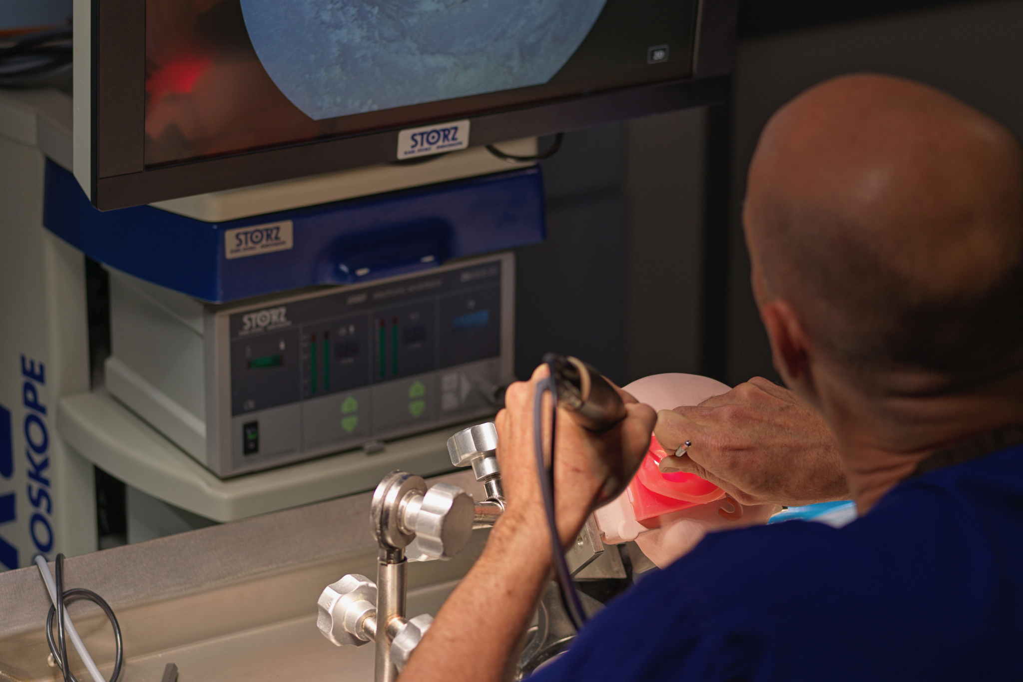
In this 360-degree video, observe a dry lab dissection in an endoscopic approach to raising a tympanomeatal flap.
Clinical Context
The dissection of a tympanomeatal flap in the context of endoscopic cholesteatoma surgery is a critical surgical step that provides access to the middle ear and lateral skull base while preserving surrounding structures. Cholesteatoma, characterised by the abnormal growth of keratinising squamous epithelium within the middle ear and mastoid, can lead to progressive bone erosion, hearing loss, facial nerve injury, and potentially life-threatening complications if not adequately managed. The minimally invasive endoscopic approach to cholesteatoma removal has gained increasing popularity due to its ability to provide enhanced visualisation of hidden recesses within the middle ear, thereby facilitating complete disease clearance while minimising morbidity.
The tympanomeatal flap dissection is an essential component of this approach, providing direct access to the middle ear space via the external auditory canal without the need for more extensive postauricular or mastoid incisions. In paediatric and adult patients alike, this step is crucial for visualising and accessing cholesteatoma in anatomically challenging areas such as the epitympanum, retrotympanum, sinus tympani, and facial recess.
The flap is typically raised after a precise circumferential incision is made in the skin of the external auditory canal, often using a 0-degree or 30-degree endoscope for optimal visualisation. The tympanomeatal flap, consisting of the canal skin and lateral tympanic membrane, is carefully elevated to expose the middle ear while avoiding injury to the tympanic annulus, chorda tympani nerve, and underlying ossicles. This meticulous dissection requires refined instrument handling and thorough knowledge of temporal bone anatomy, both of which are enhanced through cadaveric training and simulation.
Dissecting a tympanomeatal flap in cholesteatoma surgery allows for complete disease assessment and removal while preserving healthy tissue and facilitating reconstructive efforts, such as tympanic membrane grafting or ossiculoplasty. The endoscopic technique reduces the need for extensive bone drilling, offers better visualisation of disease in hidden areas, and may result in faster recovery and improved cosmetic outcomes compared to traditional microscopic approaches.
In the broader clinical context, mastering tympanomeatal flap dissection is an essential skill for otologic surgeons performing endoscopic ear surgery. It enables safe, effective management of cholesteatoma and contributes to achieving the surgical goals of eradicating disease, preserving or restoring hearing, and preventing recurrence or complications.
Learning Outcomes
- Observe an endoscopic dissection of a tympanomeatal flap.
- Understand the 3D anatomical skills required to complete this procedure.
- Observe the key steps involved in the procedure and the equipment needed to safely perform this.
External Resources