
Drilling Cortical Mastoidectomy
ENT AnatomyUse this resource in conjunction with your real-world training

Experience Summary
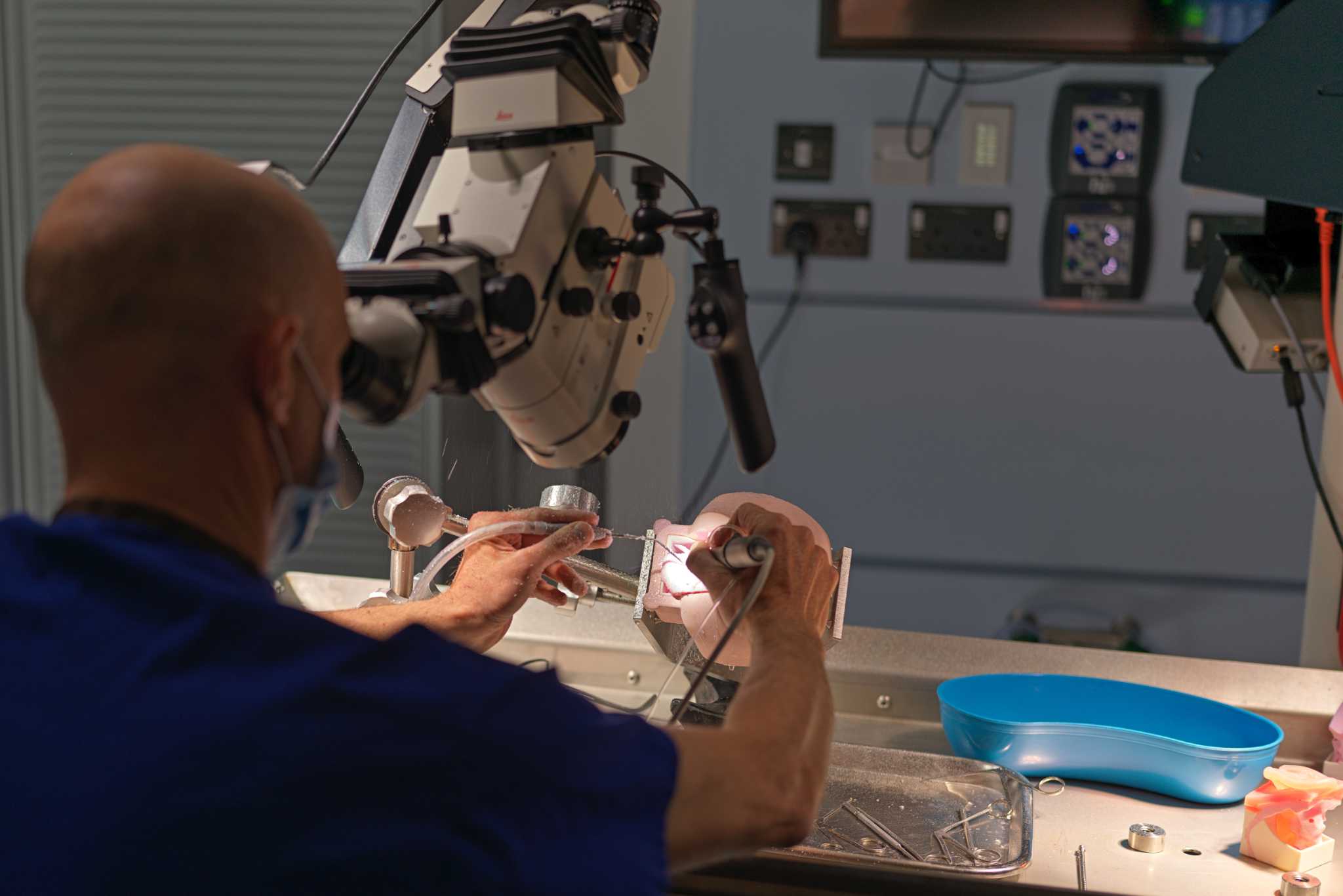
In this 360-degree video, observe a dry lab dissection of a cortical mastoidectomy procedure for cholesteatoma surgery.
Clinical Context
Drilling a cortical mastoidectomy is a fundamental surgical technique performed to access and treat disease within the mastoid portion of the temporal bone. This procedure involves removing the outer (cortical) layer of the mastoid bone to enter the mastoid air cell system, providing access to the middle ear, antrum, and deeper temporal bone structures. It is a core component of many otologic surgeries and plays a crucial role in the management of chronic ear disease, including cholesteatoma, chronic suppurative otitis media, mastoiditis, and in some cases, lateral skull base pathology.
Cortical mastoidectomy is often indicated in patients with chronic otitis media when medical management fails, and disease extends into the mastoid air cells. It is also routinely performed in conjunction with tympanoplasty for cholesteatoma removal or to drain an acute mastoiditis abscess. The primary aims of cortical mastoidectomy are to eradicate infected or diseased air cells, improve ventilation of the middle ear system, and establish safe surgical access to critical anatomical structures such as the facial nerve, sigmoid sinus, and middle cranial fossa dura.
The drilling of a cortical mastoidectomy requires detailed anatomical knowledge of the temporal bone and precision in surgical technique to avoid injury to vital structures. The surgeon begins by making a postauricular incision and elevating soft tissues to expose the mastoid cortex. Using a high-speed drill with appropriate burrs, the cortical bone is carefully removed, and the underlying mastoid air cells are opened. The dissection proceeds towards key landmarks, including the tegmen (roof of the mastoid), sigmoid sinus (posterior boundary), and the external auditory canal (anterior boundary).
In the context of cholesteatoma surgery, cortical mastoidectomy facilitates complete disease clearance, particularly when cholesteatoma extends into the mastoid antrum or attic. It also provides a route for safe identification of the lateral semicircular canal, facial nerve, and other vital structures. Drilling is performed under constant irrigation to prevent thermal injury, and meticulous technique is essential to avoid complications such as dural exposure, sigmoid sinus injury, or facial nerve damage.
In both adult and paediatric patients, cortical mastoidectomy remains a critical surgical approach for managing complex ear disease. It allows for effective disease eradication, improved middle ear ventilation, and long-term preservation of hearing and ear function, while preventing potentially life-threatening complications.
Learning Outcomes
- Observe a cortical mastoidectomy with a microscope.
- Understand the 3D anatomical skills required to complete this procedure.
- Observe the key steps involved in the procedure and the equipment needed to safely perform this.
External Resources