
Native Biopsy - 1
Renal BiopsyUse this resource in conjunction with your real-world training

Experience Summary
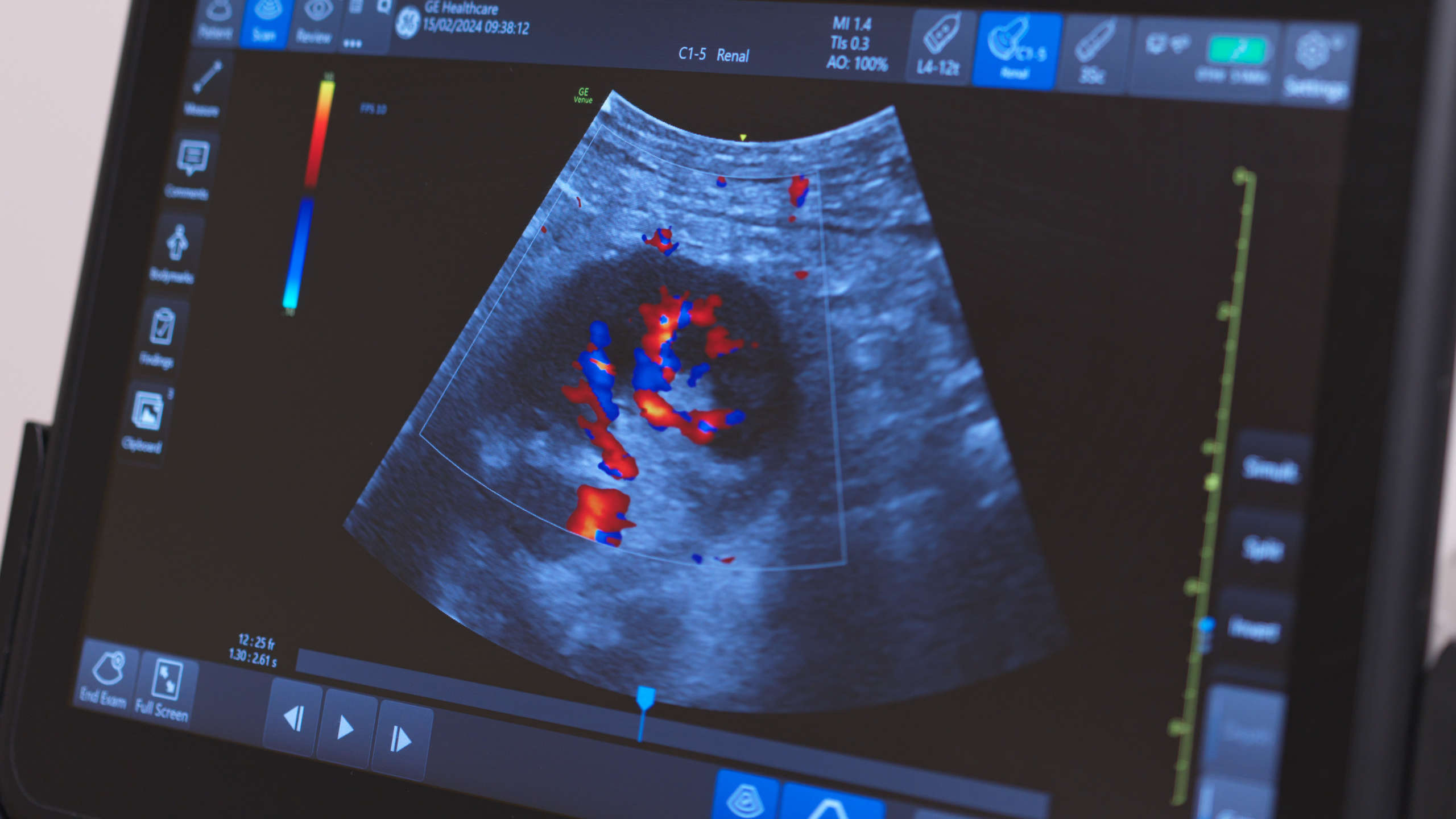
In this 360-degree video, observe a renal biopsy on a real-life patient with a native kidney.
A renal biopsy is a critical diagnostic procedure used to obtain kidney tissue for histopathological analysis. It plays a key role in evaluating unexplained renal dysfunction, proteinuria, hematuria, and suspected glomerular disease, or in monitoring transplant rejection. Performing the biopsy under ultrasound guidance enhances safety, accuracy, and efficiency, making it the standard method in most settings.
Clinical Context
Indications
A kidney biopsy is indicated when the cause of kidney impairment is uncertain and the result is likely to change management. Common indications include:
- Nephrotic or nephritic syndrome
- Acute kidney injury (AKI) of unclear origin
- Systemic diseases with renal involvement (e.g., lupus nephritis, vasculitis)
- Monitoring or diagnosing transplant dysfunction
Contraindications and Pre-Procedural Assessment
Absolute contraindications include:
- Uncorrected bleeding diathesis
- Uncontrolled hypertension
- Active infection at the biopsy site
- Solitary or small kidney (relative contraindication)
Prior to the biopsy, a thorough clinical history and assessment must be undertaken, including review of:
- Anticoagulant or antiplatelet use
- Coagulation profile (INR, APTT, platelet count)
- Blood pressure control
- Imaging to confirm anatomy and feasibility of biopsy
Patients must be counselled regarding risks such as bleeding, pain, arteriovenous fistula formation, or, rarely, loss of renal function. Consent should include explanation of benefits, risks, and post-procedural care.
Procedure Overview
The procedure is typically performed in a prone position under local anaesthesia (or lateral decubitus for transplant kidneys). Ultrasound is used to identify the lower pole of the kidney, where there is less vascular density, reducing bleeding risk.
Steps include:
- Real-time ultrasound locates the kidney and identifies an appropriate biopsy site.
- Skin is sterilised and local anaesthetic is administered.
- A spring-loaded biopsy needle is introduced under ultrasound guidance to obtain 2–3 core samples.
- Samples are checked immediately to ensure adequacy.
Post-Procedure Care
Patients are monitored for 6–24 hours, with regular observation of:
- Vital signs
- Urine output and hematuria
- Hemoglobin level to detect bleeding
Ultrasound may be repeated if bleeding is suspected. Patients are advised to avoid strenuous activity for several days.
Learning Outcomes
- Observe a full, real life renal biopsy
- Understand the equipment involved and personnel required in performing a renal biopsy
- Begin to understand how the ultrasound images guide the procedure of a renal biopsy
External Resources