
Erector Spinae Plane Block & catheter insertion
Regional AnaesthesiaUse this resource in conjunction with your real-world training

Experience Summary
In this 360-degree video, observe the anaesthetic team perform an erector spinae block and catheter insertion.
These films have been created for training purposes with individual roles played by medical professionals and/or mannequins.
No actual patients have been used in the videos.
The processes and the procedures follow the Royal Berkshire NHS Foundation trust guidelines at the time of creation.
Clinical Context
The erector spinae plane (ESP) block is a relatively recent regional anaesthetic technique used for both acute and chronic pain management. It involves the injection of local anaesthetic into the fascial plane deep to the erector spinae muscle, typically at the thoracic level, to achieve multi-dermatomal analgesia. The addition of a catheter allows for continuous infusion, extending the duration of analgesia. This technique is gaining popularity due to its simplicity, safety profile, and effectiveness in a wide range of thoracic and abdominal surgical procedures.
The ESP block is commonly indicated for post-operative pain control following procedures such as thoracotomies, rib fractures, breast surgeries, abdominal surgeries, and spinal surgeries. It is considered an alternative to neuraxial blocks like thoracic epidurals, particularly in patients with contraindications such as coagulopathy or spinal deformities. The block achieves analgesia by spreading local anaesthetic to the dorsal and ventral rami of the spinal nerves and possibly into the paravertebral space.
ESP blocks are especially useful in enhanced recovery after surgery pathways, contributing to reduced opioid consumption, improved respiratory function, and greater patient satisfaction.
Procedure
The ESP block is typically performed under ultrasound guidance in a sterile environment. The patient is positioned either sitting or in the lateral decubitus position.
- Identification of landmarks: A high-frequency linear or low-frequency curvilinear ultrasound probe is placed in a parasagittal orientation 2–3 cm lateral to the spinous process, usually at the T5–T7 level for thoracic procedures.
- Ultrasound anatomy: The key sonographic structures include the trapezius, rhomboid major, erector spinae muscle, and the transverse process.
- Needle insertion: An echogenic needle is advanced in-plane in a cranial-to-caudal direction until the tip reaches the fascial plane between the erector spinae muscle and the transverse process.
- Injection: After negative aspiration, 20–30 mL of local anaesthetic is injected, with visualisation of fluid lifting the erector spinae muscle.
- Catheter placement: If continuous analgesia is desired, a catheter is threaded through the needle into the fascial plane and secured externally for infusion.
Learning Outcomes
- Observe the procedure of performing an erector spinae plane block.
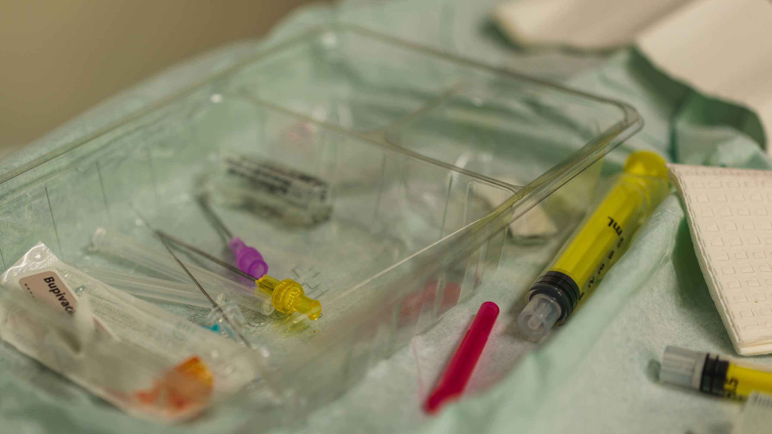
- Understand the equipment required to perform an erector spinae plane block.
- Understand the key safety steps in performing an erector spinae plane block.
External Resources