
Pleural Tap
Acute Care Common Stem (ACCS)Use this resource in conjunction with your real-world training

Experience Summary
In this 360-degree video, observe the procedure of performing a pleural aspiration of a large right-sided pneumothorax.
Clinical Context
Pleural aspiration is a key intervention in the management of a large primary spontaneous pneumothorax in clinically stable patients, particularly in line with British Thoracic Society (BTS) and NICE guidelines. In patients with minimal symptoms and no underlying lung disease, aspiration can often re-expand the lung and avoid the need for chest drain insertion. It is especially suitable when the pneumothorax occupies more than 2 cm at the level of the hilum on chest X-ray but the patient is haemodynamically stable and not in respiratory distress.
Pleural aspiration serves both diagnostic and therapeutic purposes, although in the context of pneumothorax, its role is primarily therapeutic—to evacuate air from the pleural space and allow the lung to re-expand. If aspiration fails (defined as persistent large pneumothorax or recurrence on repeat imaging), a chest drain may then be required.
Steps of Performing a Pleural Aspiration for Pneumothorax:
- Pre-procedure Assessment and Consent:
- Confirm the diagnosis and size of the pneumothorax with imaging (usually chest X-ray or point-of-care ultrasound).
- Ensure the patient is hemodynamically stable (normal BP, no hypoxia or tachypnoea).
- Obtain informed consent, explaining risks such as failure of the procedure, infection, bleeding, or rare complications like re-expansion pulmonary oedema.
- Patient Positioning:
- Position the patient sitting upright with their arms supported forward on a table or pillow to expose the lateral chest wall.
- Monitor vital signs and administer supplemental oxygen.
- Site Selection and Preparation:
- Identify the insertion site, typically the 2nd intercostal space in the midclavicular line or 5th intercostal space in the mid-axillary line, depending on operator preference and local protocols.
- Clean the skin with antiseptic and drape the area.
- Infiltrate local anaesthetic into the skin, subcutaneous tissue, and down to the pleural space, aiming just above the rib to avoid the neurovascular bundle.
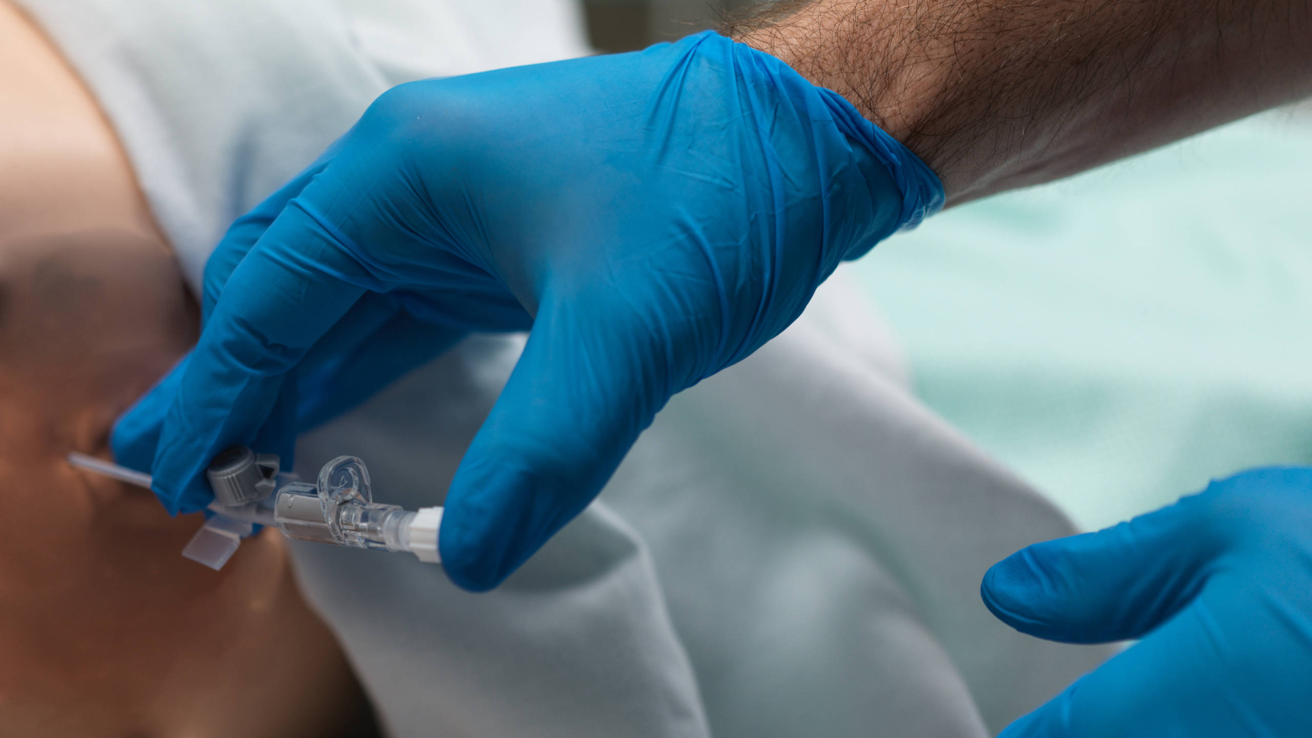
- Aspiration Technique:
- Insert a large-bore cannula into the pleural space until air is freely aspirated.
- Attach a 3-way tap and 50 mL syringe, and gradually aspirate up to 2.5–3.0 litres of air or until resistance is felt or symptoms resolve.
- Post-procedure Care:
- Remove the cannula and apply a sterile dressing.
- Reassess the patient clinically and repeat chest X-ray after 15–30 minutes.
- If the lung has re-expanded, no further intervention is needed; if not, escalate to chest drain insertion.
Learning Outcomes
- Observe the procedure of pleural aspiration in the context of a pneumothorax.
- Understand the procedural steps of pleural aspiration in the context of a pneumothorax.
- Understand the equipment required in pleural aspiration.
- Understand the key safety points in pleural aspiration.
External Resources