
CVC Line Placement
Anaesthetic Critical IncidentsUse this resource in conjunction with your real-world training

Experience Summary
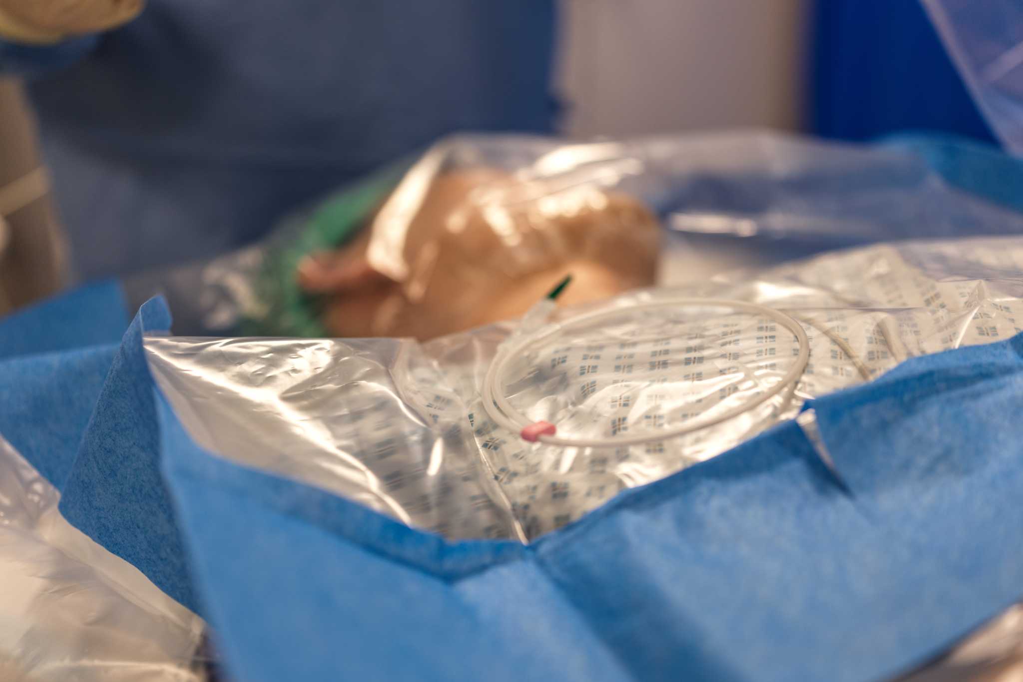
In this experience, watch a simulated central venous cannulation procedure.
Clinical Context
Central venous catheter (CVC) insertion is a common invasive procedure in anaesthetic and critical care practice, used to obtain reliable venous access and facilitate advanced monitoring and therapy. It is typically performed in operating theatres, intensive care units, or emergency settings. The clinical context for CVC insertion includes patients requiring vasoactive infusions, difficult peripheral access, rapid fluid resuscitation, or administration of irritant drugs such as chemotherapy or hyperosmolar solutions (e.g. total parenteral nutrition). It is also indicated for central venous pressure monitoring, though this is used less frequently in modern practice. Common insertion sites include the internal jugular, subclavian, and femoral veins, each with specific advantages and risks.
Procedural Steps of CVC Insertion
Preparation is essential to ensure safety and success. The procedure should be performed with full aseptic technique, including hand hygiene, sterile gown, gloves, drapes, and skin preparation with chlorhexidine. Appropriate monitoring (ECG, pulse oximetry, blood pressure) should be in place, and resuscitation equipment readily available. Ultrasound guidance is now considered standard of care, particularly for internal jugular access, as it improves success rates and reduces complications. The procedure is most commonly performed using the Seldinger technique. After identifying the target vein with ultrasound, local anaesthetic is infiltrated into the skin and subcutaneous tissues. A needle is advanced under ultrasound guidance into the vein, and correct placement is confirmed by free aspiration of venous blood. A guidewire is then passed through the needle into the vessel, and the needle is removed while maintaining wire position. A small skin incision may be made to facilitate passage of the dilator. The tract is then gently dilated, and the central venous catheter is advanced over the guidewire into the vessel. The guidewire is removed, and all lumens are aspirated and flushed to confirm patency. The catheter is then secured with sutures or a fixation device and covered with a sterile dressing. Correct positioning must be confirmed, typically with a chest X-ray for internal jugular or subclavian lines, to ensure the tip lies in the lower superior vena cava and to exclude complications such as pneumothorax. Ultrasound or ECG-guided tip confirmation may also be used.
Complications of CVC Insertion
Complications include arterial puncture, haematoma, pneumothorax, infection, thrombosis, and arrhythmias during wire insertion. Careful technique and ultrasound guidance minimise these risks.
Learning Outcomes
- Observe the procedure of CVC insertion on a simulated patient.
- Understand the procedural steps of CVC insertion.
- Understand the relevant anatomy to CVC insertion.
- Understand the potential complications of CVC insertion.
External Resources