
Venous Thromboembolism (VTE) Risk Assessment
Maternal MedicineUse this resource in conjunction with your real-world training

Experience Summary
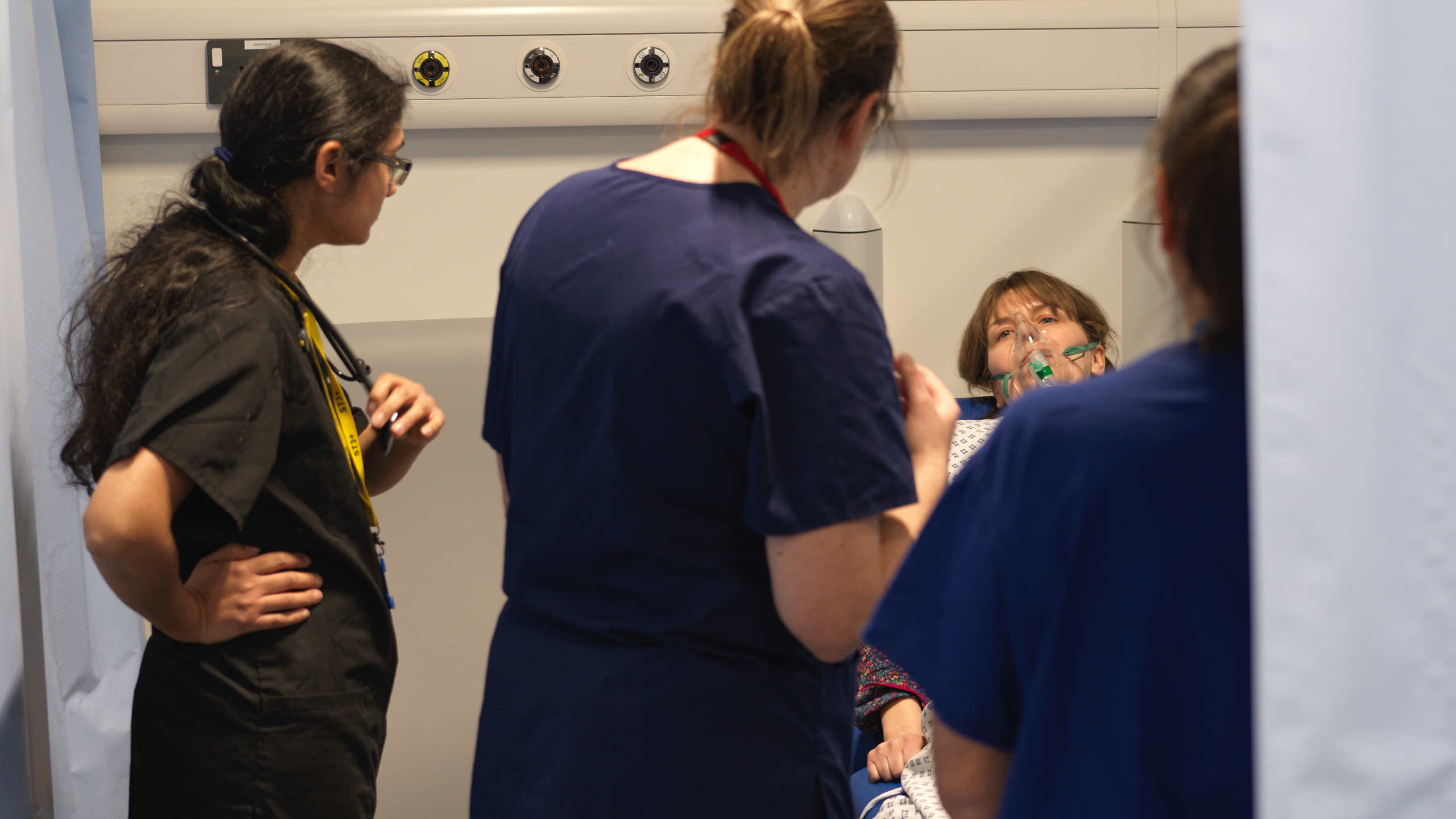
In this 360-degree video, observe a patient attending for a booking appointment and venous thromboembolism (VTE) risk assessment. Observe how the communication and assessment at the booking appointment can impact later clinical outcomes for the patient.
Clinical Context
Pulmonary embolism (PE) is a leading cause of maternal morbidity and mortality, accounting for a significant proportion of maternal deaths worldwide. Pregnancy is a recognised hypercoagulable state due to physiological changes that promote clot formation, intended to reduce the risk of haemorrhage during childbirth. However, these changes also increase the risk of VTE, including deep vein thrombosis (DVT) and PE.
The risk of PE is estimated to be 4-5 times higher in pregnant women compared to non-pregnant women of the same age, with the highest risk occurring in the postpartum period, particularly the first six weeks after delivery.
Risk Factors for PE in Pregnancy
- Personal or family history of VTE.
- Known thrombophilia (e.g., Factor V Leiden, antiphospholipid syndrome).
- Obesity (BMI > 30).
- Advanced maternal age (> 35 years).
- Smoking.
- Immobility (e.g., prolonged bed rest, post-surgery).
- Caesarean section.
- Multiple pregnancy.
- Pre-eclampsia.
Initial Risk Assessment and Clinical Presentation
Early identification of PE in pregnancy is challenging, as symptoms such as breathlessness, tachycardia, and leg swelling can overlap with normal physiological changes of pregnancy. High clinical suspicion is essential, particularly in women with risk factors.
Typical presenting features include:
- Sudden onset dyspnoea.
- Pleuritic chest pain.
- Tachypnoea.
- Tachycardia.
- Haemoptysis (less common).
- Signs of DVT (e.g., unilateral leg swelling, tenderness).
Assessment
Initial assessment follows an ABCDE approach to stabilise the patient and identify red flags. Oxygen saturation, heart rate, respiratory rate, and blood pressure should be monitored closely.
Diagnostic tools include:
- Wells Score and D-dimer testing are not validated for pregnancy.
- Lower limb Doppler ultrasound for suspected DVT.
- CT Pulmonary Angiogram (CTPA) or Ventilation-Perfusion (V/Q) scan for suspected PE, balancing maternal and fetal radiation exposure. Current guidelines recommend CTPA as first-line imaging in most cases.
Management
If PE is suspected, treatment should not be delayed while awaiting imaging. **Low Molecular Weight Heparin (LMWH)**is the first-line treatment as it does not cross the placenta and is considered safe in pregnancy. Dosing is based on maternal weight and continued throughout pregnancy and for at least six weeks postpartum, or until at least three months of anticoagulation is completed.
Multidisciplinary input from obstetrics, haematology, and respiratory or critical care teams is essential for optimal management.
Learning Outcomes
- Reflect on the communication skills during the booking consultation.
- Understand the risk factors for VTE in this patient.
- Understand the symptoms of PE.
- Understand the investigation options for VTE in pregnant patients.
- Understand the treatment options for VTE in pregnant patients.
External Resources