
Normal upper airway with an established tracheostomy
Tracheostomy and Airway ManagementUse this resource in conjunction with your real-world training

Experience Summary
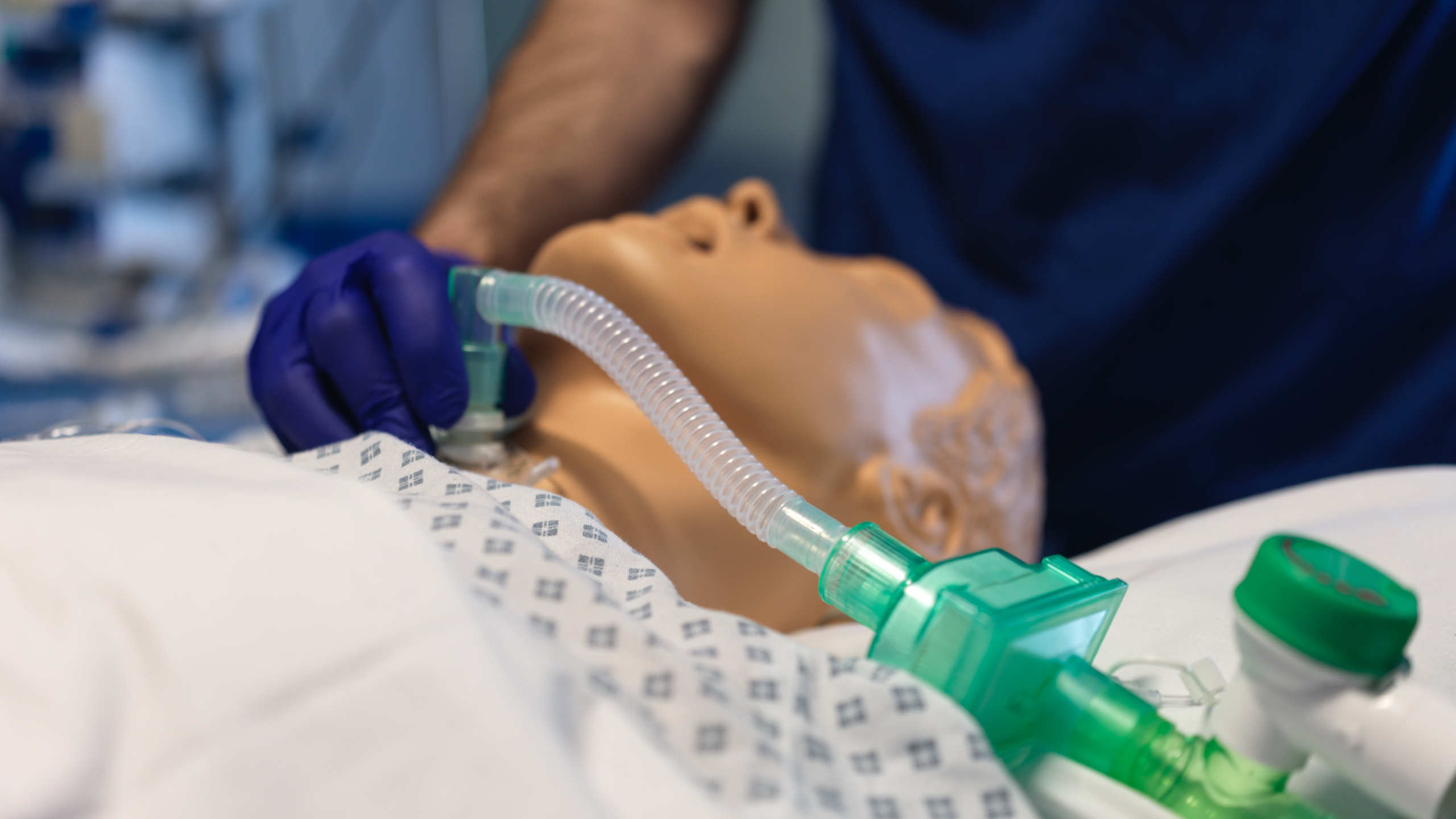
This experience demonstrates an overview of the key steps in managing a tracheostomy emergency. The experience is presented from the point-of-view of an observer, so you can see all of the key steps, vital signs monitors, equipment and the human factors and interactions between the team.
The team explain their actions and work their way through airway assessment, tracheostomy assessment, basic airway management, and management of the blocked tracheostomy. The patient had an established (1 year old) surgical tracheostomy and a normal upper airway. The team manage the situation by removing the tracheostomy (after establishing it is blocked or displaced) and directly re-inserting a new tracheostomy tube, as this is the easiest airway.
Clinical Context
A blocked tracheostomy tube is a life-threatening emergency that can rapidly lead to hypoxia and cardiac arrest if not promptly identified and managed. Patients with tracheostomies are commonly encountered in intensive care, postoperative settings, and long-term care facilities. Tracheostomy tubes may become obstructed due to thick secretions, blood clots, kinking of the tube, or granulation tissue formation. High-risk patients include those with poor cough effort, inadequate humidification, or underlying respiratory disease.
Clinically, a blocked tracheostomy should be suspected when a patient with a tracheostomy becomes acutely distressed, exhibits increased work of breathing, reduced oxygen saturations, or silent chest on auscultation. Capnography, if available, may show absent or reduced end-tidal CO₂. There may be visible signs of respiratory distress, accessory muscle use, cyanosis, or altered consciousness.
Management follows an immediate, structured approach. First, call for help and apply high-flow oxygen via a tracheostomy mask or a facemask over both the mouth and stoma. Assess for tube patency by attempting to pass a suction catheter. Inability to pass the catheter confirms obstruction.
If blocked, remove the inner cannula (if present), as this is a common site of obstruction. Reassess breathing. If the patient remains compromised, consider removing the entire tracheostomy tube and providing oxygen via the stoma or upper airway, depending on the patient’s ventilation route and clinical status.
If the patient is known to be able to breathe via the upper airway, oral ventilation with a bag-valve mask may be attempted. In cuffed, non-fenestrated tracheostomies, upper airway breathing may not be possible, and the stoma must be used.
Definitive management includes suctioning, replacing the tube, or re-establishing airway access with help from experienced personnel. Familiarity with emergency tracheostomy algorithms, regular staff training, and clear documentation of airway plans are critical for safe, effective management.
Learning Outcomes
- Ventilation of the stoma via a paediatric facemask and a supraglottic airway device.
- Re-insertion of the tracheostomy tube directly into the established stoma.
External Resources